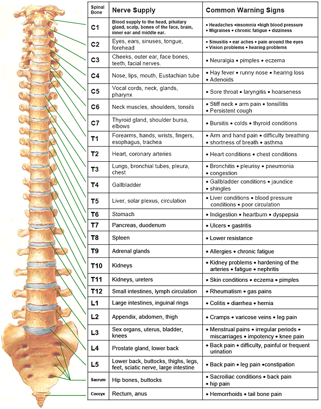
Web the nerve roots are numbered based on their location in the spinal cord, with the first cervical nerve root (c1) located at the top of the spinal cord near the base of the brain, and the last sacral nerve root (s5) located at the bottom. These nerves are essential for transmitting sensory signals to the brain and for carrying motor commands from the brain to muscles. Web how to use the spinal nerve chart: Web background bilateral diaphragmatic dysfunction can lead to dyspnea and recurrent respiratory failure. Many of the nerves of the peripheral nervous system, or pns, branch out from the.
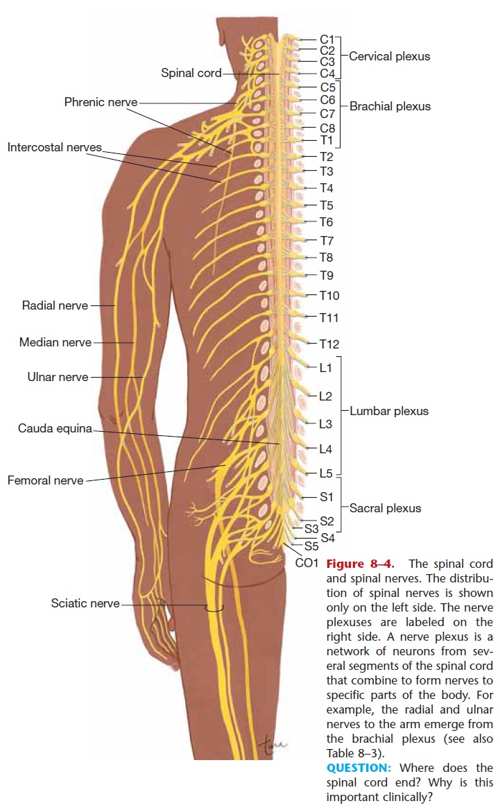
The spinal cord carries messages back and forth between the brain and the nerves that run throughout the body. Web background bilateral diaphragmatic dysfunction can lead to dyspnea and recurrent respiratory failure. Web below is a chart that outlines the main functions of each of the spine nerve roots: Eight cervical spinal nerve pairs, 12 thoracic pairs , five lumbar pairs, five sacral pairs, and one coccygeal. Each one is named after the vertebra beneath it, except the c8 nerves, which are above the t1 vertebra.
The peripheral nerves are responsible for sensations and muscle movements. Web to understand this intricate region, we will consider the bony structures first, and then discuss the ligaments, nerves, and musculature that are associated with this region of the spinal column, concluding with some clinical implications of damage to some of these structures. Web the central nervous system is made up of the brain and spinal cord: The worst place to get a tattoo and notable seconds. Web these relay motor (movement), sensory (sensation), and autonomic (involuntary functions) signals between the spinal cord and other parts of the body.
Spinal Nerve Function Anatomical Chart Anatomy Models and Anatomical

Spinal Nerves
Printable Spinal Nerve Chart
Printable Spinal Nerve Chart Printable World Holiday

The Spine and Spinal Nerves Poster Clinical Charts and Supplies
Nerve Chart Hunter Chiropractic Wellness Centre

Anatomical Charts and Posters Anatomy Charts Spinal Nerves
Important Nerves in the Body and What They Do NorthEast Spine and
Spinal Nerve Chart

Spinal Nerves Anatomical Chart Spine and Cranial Nervous System

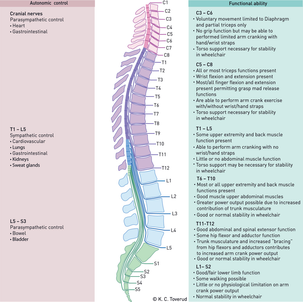
Web your spine is a complex structure of small bones, cushioning disks, nerves, joints, ligaments and muscles. [1] [2] these are grouped into the corresponding cervical, thoracic, lumbar, sacral and coccygeal regions of the spine. Many of the nerves of the peripheral nervous system, or pns, branch out from the. On the chart below you will see 4 columns (vertebral level, nerve root, innervation, and possible symptoms). This chart illustrates spinal nerves, cranial nerves and diagrams the portion of the thoracic spinal cord with spinal nerves. Web below is a chart that outlines the main functions of each of the spine nerve roots: Web spinal nerves are mixed nerves that emerge from the spinal cord and carry both motor and sensory information between the spinal cord and various parts of the body. Each one is named after the vertebra beneath it, except the c8 nerves, which are above the t1 vertebra. 20 (w) x 26 (h) your trusted supplier for spinal nerves anatomical charts. The worst place to get a tattoo and notable seconds. The vertebral column’s most important physiologic function is protecting the spinal cord, which is the main avenue for communication between the. Web the nerve roots are numbered based on their location in the spinal cord, with the first cervical nerve root (c1) located at the top of the spinal cord near the base of the brain, and the last sacral nerve root (s5) located at the bottom. Web to understand this intricate region, we will consider the bony structures first, and then discuss the ligaments, nerves, and musculature that are associated with this region of the spinal column, concluding with some clinical implications of damage to some of these structures. A dermatome is an area of skin supplied by a single spinal nerve. Web the central nervous system is made up of the brain and spinal cord:
This Chart Illustrates Spinal Nerves, Cranial Nerves And Diagrams The Portion Of The Thoracic Spinal Cord With Spinal Nerves.
Eight cervical spinal nerve pairs, 12 thoracic pairs , five lumbar pairs, five sacral pairs, and one coccygeal. Web the nerve roots are numbered based on their location in the spinal cord, with the first cervical nerve root (c1) located at the top of the spinal cord near the base of the brain, and the last sacral nerve root (s5) located at the bottom. Thoracic spinal nerves are not part of any plexus, but give rise to the intercostal nerves directly. For the most part, the spinal nerves exit the vertebral canal through the intervertebral foramen below their corresponding vertebra.
Each Spinal Nerve Is Supplied By 2 Nerve Roots.
Both the brain and the spinal cord are protected by bone: Web below is a chart that outlines the main functions of each of the spine nerve roots: Web there are 8 pairs of spinal nerves in the cervical spine, labeled c1 to c8. Web there are 5 pairs of spinal nerves in the lumbar spine, labeled l1 to l5.
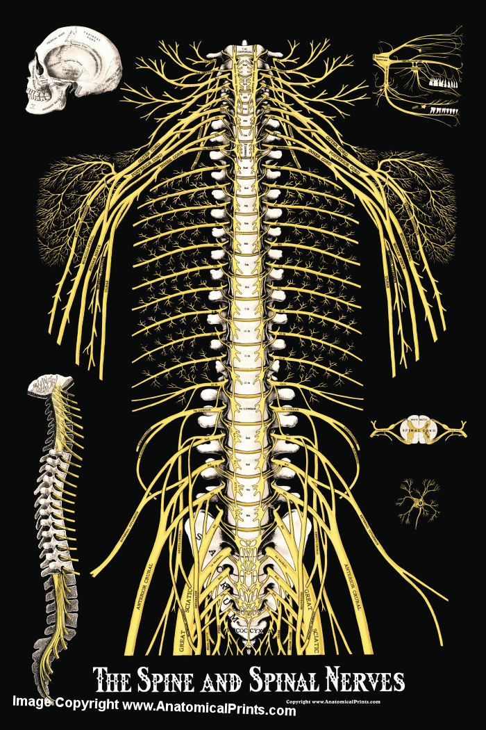
Each Of These Nerves Branches Out From The Spinal Cord, Dividing And Subdividing To Form A Network Connecting The Spinal Cord To Every Part Of The Body.
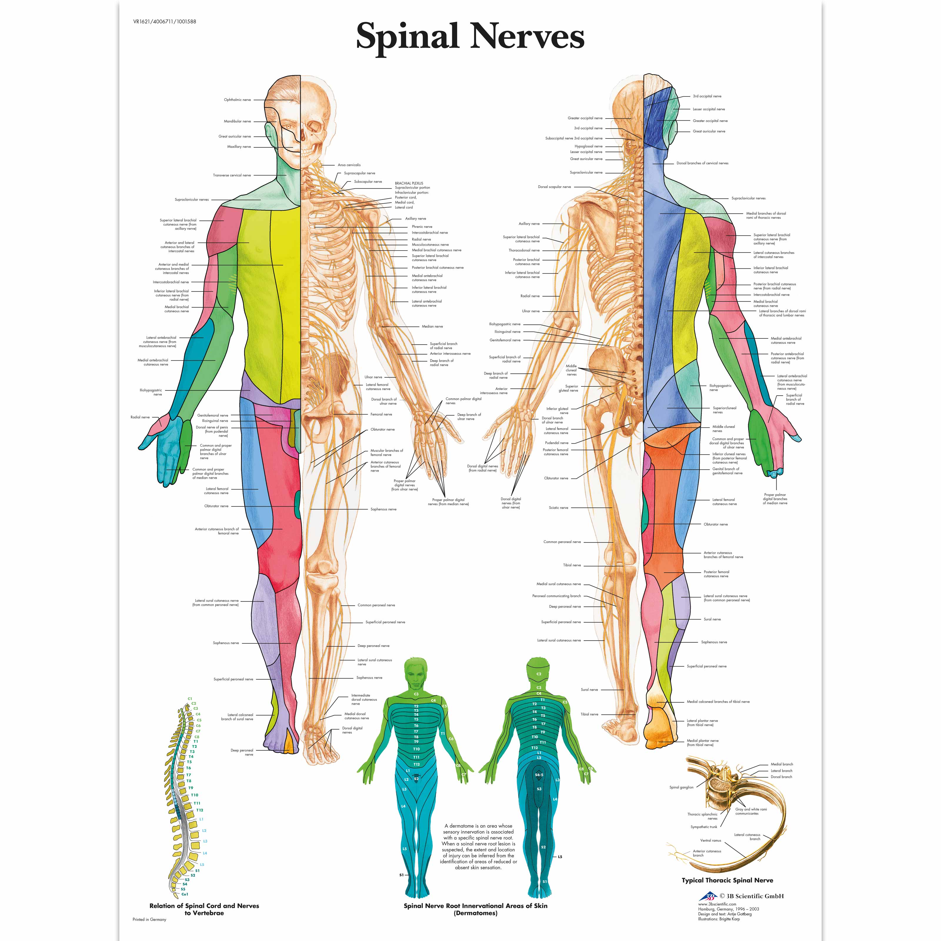
The spine is a major part of the nervous system and has many sensory nerves. Web these 31 pairs of spinal nerves are like the backbone of your peripheral nervous system, making sure your brain and body stay in sync. A dermatome is an area of skin supplied by a single spinal nerve. Web a spinal nerve chart provides a visual reference to help memorize the vertebral levels, sensory pathways, and motor functions of the network of nerves that transmit signals between the central nervous system and periphery.
The Peripheral Nerves Are Responsible For Sensations And Muscle Movements.
Spinal cord segments, cutaneous distribution of spinal nerves and dermal segmentation are also shown. Web learn how spinal nerve roots function, and the potential symptoms of spinal nerve compression and pain in the neck and lower back. The spinal cord begins at the base of the brain and extends into the pelvis. Web in the human body there are 31 pairs of spinal nerves, one on each side of the vertebral column.